SPECT-CT and PET-CT
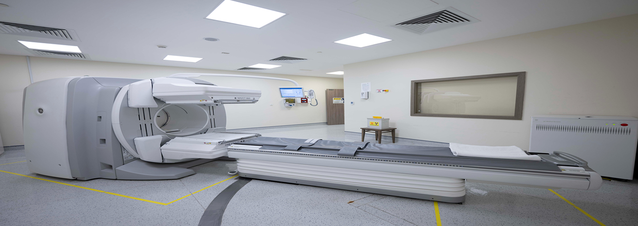
The SPECT-CT and PET-CT imaging technologies at Burjeel Cancer Institute provide advanced diagnostic tools to detect, stage, and monitor various cancers. These hybrid imaging systems combine nuclear medicine with computed tomography (CT), allowing for highly accurate detection of cancerous activity and providing detailed anatomical information. These technologies are essential in guiding treatment decisions, tracking cancer progression, and assessing the response to therapies.
Key Features
Both SPECT-CT and PET-CT offer several features that make them invaluable in cancer diagnostics and management
Early Cancer Detection
PET-CT, in particular, detects cancerous changes at the molecular level, allowing for earlier diagnosis and improved outcomes.
Functional and Anatomical Imaging
Both modalities allow physicians to view metabolic changes in cells and precise anatomical details in a single scan, enhancing diagnostic accuracy.
PET-CT (Positron Emission Tomography-Computed Tomography):
- Provides highly sensitive imaging by combining PET and CT scans, offering detailed images of cancerous lesions and metabolic activity.
- Often used to detect and monitor cancers such as lung cancer, lymphoma, and colorectal cancer.
SPECT-CT (Single Photon Emission Computed Tomography-Computed Tomography):
- Combines nuclear medicine and CT to provide both functional and anatomical imaging, making it useful for detecting cancerous activity in bones, organs, and soft tissues.
- Used for detecting bone metastases, thyroid cancer, and other cancers with metabolic activity.
Cancers Diagnosed and Monitored with SPECT-CT and PET-CT
These imaging systems are used to diagnose, stage, and monitor a wide range of cancers, including:
- Lung cancer
- Breast cancer
- Lymphoma (Hodgkin's and non-Hodgkin's)
- Colorectal cancer
- Prostate cancer
- Thyroid cancer
- Melanoma
- Head and neck cancer
- Bone metastases (secondary cancer spread to bones)
- Neuroendocrine tumors
- Other solid tumors and metastatic cancers
Benefits of SPECT-CT and PET-CT
These advanced imaging systems provide numerous benefits to patients and physicians:
Accurate Diagnosis
PET-CT and SPECT-CT offer a comprehensive view of cancer activity and anatomical structures, allowing for more accurate diagnosis and staging of cancer.
Early Detection
PET-CT can detect metabolic changes in cells before structural changes are visible, allowing for earlier diagnosis and improved treatment outcomes.
Treatment Planning
The detailed images provided by these scans guide radiation oncologists and surgeons in planning precise treatments, such as radiation therapy or surgical tumor removal.
Monitoring Treatment Response
Both technologies are invaluable for assessing how well a treatment is working by monitoring changes in cancerous activity, helping to adjust treatment plans if necessary.
Minimally Invasive
Both imaging techniques are non-invasive and painless, offering patients a safe way to undergo comprehensive cancer diagnostics.

Our Approach to Imaging and Diagnosis
At Burjeel Cancer Institute, SPECT-CT and PET-CT are integral to our comprehensive approach to cancer care:
Multidisciplinary Collaboration
Our radiologists, nuclear medicine physicians, oncologists, and surgeons collaborate to interpret imaging results and develop personalized treatment plans.
Personalized Diagnostic Plans
Every patient's imaging and diagnostic needs are tailored based on their type of cancer, medical history, and treatment goals.
Advanced Imaging Techniques
We utilize the latest PET-CT and SPECT-CT technology to ensure the highest level of accuracy in cancer diagnosis and treatment planning.
Focus on Precision
These imaging technologies provide precise details about the location, size, and spread of tumors, allowing for targeted and effective treatment.

Patient Journey
Patients undergoing SPECT-CT or PET-CT imaging at Burjeel Cancer Institute can expect a streamlined, supportive experience:
Initial Consultation
A thorough consultation with a physician to determine the appropriate imaging study based on the patient's type of cancer and diagnostic needs.
Imaging Preparation
Patients are guided through the process of preparing for a PET-CT or SPECT-CT scan, including any dietary restrictions or medications required before the scan.
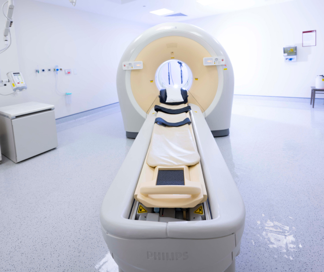
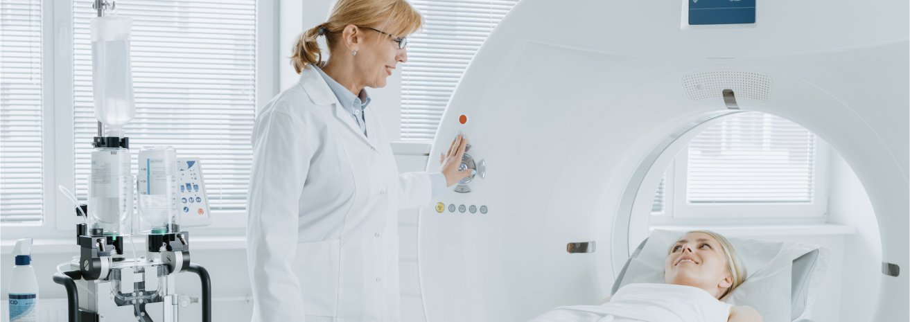
Imaging Procedure
The scan is conducted in a comfortable environment, with the patient lying still while the imaging machine takes detailed pictures.
Image Analysis
The scan results are reviewed by a specialized team, and a comprehensive report is shared with the patient's care team to guide treatment decisions.
Post-Imaging Follow-Up
Patients receive detailed feedback on the results and any changes to their treatment plan, with follow-up scans scheduled as needed to monitor progress.
