3D Mammography System

The 3D Mammography System at Burjeel Cancer Institute offers advanced breast cancer screening and diagnostic capabilities, providing clearer and more accurate imaging than traditional mammograms. Also known as Digital Breast Tomosynthesis (DBT), 3D mammography captures multiple images of the breast from different angles, creating a detailed, three-dimensional image that allows for better detection of breast abnormalities. This cutting-edge technology is particularly useful for detecting breast cancer in its early stages, even in women with dense breast tissue.
Key Features
The 3D Mammography System offers numerous features that improve the accuracy of breast cancer detection:
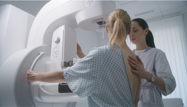
Comfortable Procedure
Comfortable Procedure: While 3D mammography uses similar compression techniques to traditional mammography, the advanced imaging technology allows for shorter scan times, enhancing patient comfort.
Improved Detection in Dense Breasts
3D mammography is especially beneficial for women with dense breast tissue, where traditional mammograms may struggle to detect abnormalities.
Reduced False Positives
By providing more detailed images, 3D mammography reduces the likelihood of false positives, minimizing the need for additional testing and reducing anxiety for patients.
Early Detection
Early Detection: The increased accuracy of 3D mammography allows for the detection of smaller tumors and subtle abnormalities, often before they are visible on standard mammograms.
Three-Dimensional Imaging
Unlike traditional 2D mammograms, 3D mammography takes multiple X-ray images of the breast from different angles, creating a detailed, layered image that provides a clearer view of breast tissue.
Cancers Detected with 3D Mammography
The 3D Mammography System is used for both breast cancer screening and diagnosis, helping to detect:
- Invasive ductal carcinoma
- Invasive lobular carcinoma
- Ductal carcinoma in situ (DCIS)
- Lobular carcinoma in situ (LCIS)
- Triple-negative breast cancer
- HER2-positive breast cancer
- Other types of early-stage and advanced breast cancers
Benefits of 3D Mammography
Patients undergoing 3D mammography benefit from its advanced capabilities in breast cancer detection:
Increased Detection Rates
3D mammography has been shown to improve breast cancer detection rates, particularly for early-stage cancers that are harder to detect with standard mammography.
Reduced Callbacks for Additional Testing
The clearer images provided by 3D mammography reduce the number of patients called back for additional imaging due to unclear or inconclusive results.
More Accurate for Dense Breasts
Women with dense breast tissue, who are at higher risk of breast cancer, benefit from the enhanced clarity and accuracy of 3D mammography, improving early detection.
Better Visualization of Breast Abnormalities
3D imaging helps to distinguish between benign and cancerous abnormalities, reducing unnecessary biopsies and further testing.
Non-Invasive and Safe
3D mammography is a non-invasive procedure that uses low-dose X-rays, making it safe and effective for routine breast cancer screening.

Our Approach to Breast Cancer Screening
At Burjeel Cancer Institute, we use the 3D Mammography System as part of our comprehensive approach to breast cancer care:
Multidisciplinary Collaboration
Our team of radiologists, breast surgeons, medical oncologists, and oncology nurses collaborates to interpret imaging results and develop personalized treatment or screening plans.
Personalized Screening Plans
We tailor breast cancer screening schedules based on each patient's risk factors, including age, family history, and breast density, ensuring timely and accurate detection.
High-Risk Screening Programs
For women at higher risk of breast cancer, we offer advanced screening programs that include 3D mammography, genetic testing, and regular follow-ups to detect cancer as early as possible.
Comprehensive Care
From screening and diagnosis to treatment and recovery, we provide compassionate, patient-centered care throughout the entire cancer care journey.
Patient Journey
Patients undergoing 3D mammography at Burjeel Cancer Institute can expect a comfortable and supportive experience:
Initial Consultation
A consultation with a physician or breast cancer specialist to assess the patient's risk factors and determine the appropriate screening or diagnostic imaging plan.
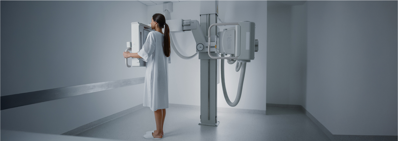
Imaging Procedure
During the 3D mammography scan, the breast is positioned and compressed similarly to a traditional mammogram. The system then takes multiple images from different angles, which are combined to create a 3D image.
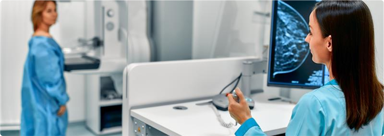
Image Analysis
The images are reviewed by a specialized radiologist, who interprets the results and provides a comprehensive report to the patient's care team.
Post-Imaging Follow-Up
Patients receive detailed feedback on the results, and any necessary follow-up imaging or biopsies are scheduled if needed. For routine screenings, patients receive regular reminders for future check-ups.
