Intraoperative MRI (iMRI)
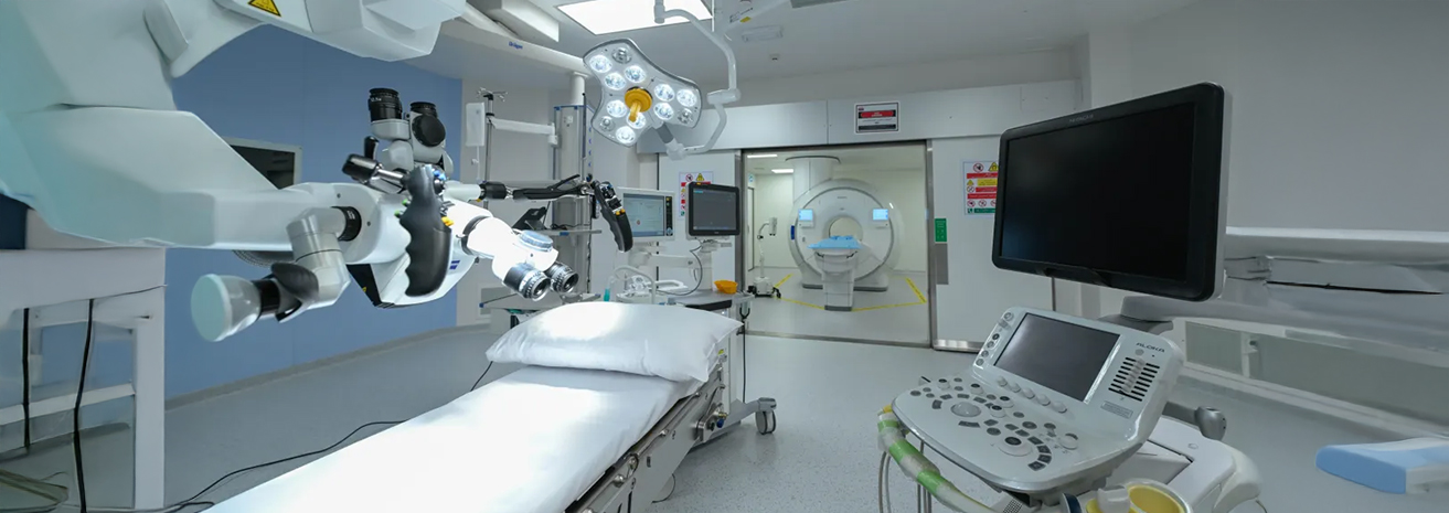
At Burjeel Cancer Institute, we offer both 1.5T and 3T MRI (Magnetic Resonance Imaging) technologies, providing advanced, high-resolution imaging for the diagnosis and monitoring of cancer. MRI is a non-invasive imaging technique that uses powerful magnetic fields and radio waves to produce detailed images of the body's internal structures. Our 3T MRI, with double the magnetic strength of the 1.5T MRI, offers exceptional clarity and precision, allowing for better visualization of small tumors and detailed anatomical features. These advanced MRI technologies are essential for accurate cancer diagnosis, treatment planning, and monitoring.
Key Features
The 1.5T and 3T MRI systems offer several advanced features for cancer diagnostics and monitoring:
Reduced Scan Times with 3T MRI
The increased magnetic strength of the 3T MRI reduces scan times, improving patient comfort while maintaining image quality.
Contrast-Enhanced MRI
Involves the use of a contrast agent to highlight blood vessels and enhance the visibility of tumors, improving diagnostic accuracy.
Diffusion-Weighted Imaging (DWI)
An MRI technique used to detect cancer by measuring the movement of water molecules in tissues, often helpful in identifying tumor spread.
Functional MRI (fMRI)
This advanced MRI technique measures brain activity by detecting changes in blood flow, often used for brain tumors or neurological complications caused by cancer.
Detailed Soft Tissue Imaging
MRI is particularly effective at visualizing soft tissues, such as the brain, liver, prostate, and breast, allowing for the accurate detection and staging of cancers.
High-Resolution Imaging
Both 1.5T and 3T MRI provide detailed, high-resolution images, with the 3T MRI offering even greater clarity, making it ideal for detecting small tumors and subtle abnormalities.
Cancers Diagnosed and Monitored with 1.5T and 3T MRI
These MRI systems are used to diagnose, stage, and monitor various types of cancers, including:
- Brain tumors
- Breast cancer
- Prostate cancer
- Liver cancer
- Pancreatic cancer
- Bone and soft tissue tumors
- Spine and spinal cord tumors
- Gynecologic cancers (e.g., ovarian and uterine cancer)
- Colorectal cancer (metastases)
- Other abdominal and pelvic tumors
Benefits of 1.5T and 3T MRI
Both 1.5T and 3T MRI systems provide a range of benefits for cancer patients:
Superior Soft Tissue Contrast
MRI offers unparalleled imaging of soft tissues, making it ideal for detecting tumors in the brain, spine, liver, and other organs where precision is critical.
Non-Invasive:
MRI scans are non-invasive and do not use ionizing radiation, making them a safe option for cancer diagnosis and follow-up imaging.
Detailed Imaging for Treatment Planning
The high-resolution images produced by MRI help guide treatment decisions, such as surgical planning or radiation therapy targeting.
Detection of Small Tumors
3T MRI is particularly effective at detecting small tumors or abnormalities that may be difficult to see on other imaging modalities, ensuring earlier diagnosis and intervention.
Functional Imaging Capabilities
Functional MRI and diffusion-weighted imaging help evaluate tumor aggressiveness, monitor response to treatment, and detect tumor recurrence.
Contrast-Enhanced Scanning
The use of contrast agents in MRI can enhance the visibility of blood vessels and tumors, improving diagnostic accuracy.
Our Approach to MRI Imaging
At Burjeel Cancer Institute, our MRI imaging services are part of a comprehensive, patient-centered approach to cancer care:
Multidisciplinary Collaboration
bOur radiologists, medical oncologists, surgeons, and radiation oncologists work together to interpret MRI results and develop personalized treatment plans for each patient.
Personalized Imaging Protocols
We tailor MRI protocols to the specific needs of each patient, ensuring that the scans provide the necessary information for diagnosis and treatment planning.
Advanced Technology
Our 3T MRI system offers the latest advancements in imaging, ensuring the highest level of accuracy for cancer diagnosis and monitoring.
Focus on Comfort and Efficiency
Our MRI systems are designed to provide a comfortable scanning experience, with reduced scan times and minimized patient discomfort, especially with the 3T MRI.

Patient Journey
Patients undergoing MRI imaging at Burjeel Cancer Institute can expect a supportive and efficient experience:
Initial Consultation
A consultation with a physician to determine the most appropriate imaging study based on the patient's cancer type and medical history.
Imaging Preparation
Patients are guided through the MRI preparation process, which may involve the use of a contrast agent to enhance image quality.
MRI Scan
The MRI scan is conducted in a comfortable environment, with patients lying still while the imaging system captures detailed images of the targeted area.
Image Analysis
The MRI images are reviewed by a specialized team, and a comprehensive report is provided to the patient's care team for diagnosis and treatment planning.
Post-Imaging Follow-Up
After the scan, the patient will receive feedback on the results and discuss any changes to their treatment plan, with follow-up scans scheduled as needed.
