3D/4K/ICG Surgical Endoscope System, Image1 S™ Rubina®
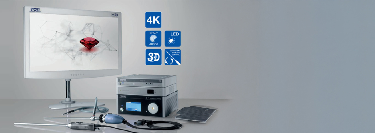
The 3D/4K/ICG Surgical Endoscope System, Image1 S™ Rubina® at Burjeel Cancer Institute represents the latest in minimally invasive surgical technology. This cutting-edge system combines high-resolution 4K imaging, 3D visualization, and near-infrared fluorescence (ICG) imaging to provide surgeons with enhanced precision and clarity during complex procedures. The integration of 3D/4K technology allows for superior depth perception, while the ICG fluorescence feature helps visualize blood flow and tissue perfusion in real-time, making it particularly valuable in oncologic surgeries where accuracy and tissue differentiation are crucial.
Key Features
The Image1 S™ Rubina® system offers numerous advanced features that enhance surgical precision and improve patient outcomes:
Enhanced Color Display
The system provides vibrant color representation and real-time visualization, allowing surgeons to differentiate between healthy and diseased tissues more easily.
Multispecialty Application
Multispecialty Application: The versatility of the system makes it suitable for a wide range of oncologic surgeries, including gastrointestinal, liver, gynecologic, and thoracic surgeries.
ICG Fluorescence Imaging
ICG Fluorescence Imaging: The system includes indocyanine green (ICG) fluorescence technology, which allows for real-time visualization of blood vessels, lymph nodes, and bile ducts, aiding in the identification of cancerous tissues and ensuring complete tumor removal.
3D Visualization
The 3D capabilities allow for enhanced depth perception, enabling surgeons to perform more precise dissection and tissue manipulation during minimally invasive procedures.
4K Ultra-High-Definition Imaging
The system delivers crystal-clear 4K UHD resolution, providing exceptional detail that helps surgeons visualize fine anatomical structures during surgery.
Conditions Treated Using the Image1 S™ Rubina® System
This advanced surgical endoscopy system is used in a variety of minimally invasive cancer surgeries, including:
- Colorectal cancer
- Liver cancer (including hepatocellular carcinoma and metastases)
- Pancreatic cancer
- Gynecologic cancers (e.g., ovarian and uterine cancer)
- Lung cancer (for thoracoscopic procedures)
- Esophageal cancer
- Gastric cancer
- Prostate cancer
- Biliary tract and gallbladder cancers
- Minimally invasive biopsies and resections
Benefits of the Image1 S™ Rubina® System
Patients undergoing surgery using the 3D/4K/ICG Surgical Endoscope System benefit from its advanced visualization and imaging capabilities:
Enhanced Tumor Visualization
The ICG fluorescence feature allows surgeons to visualize blood flow and tissue perfusion in real-time, ensuring more accurate tumor resections and reducing the risk of leaving cancerous cells behind.
Superior Depth Perception
The 3D technology provides enhanced depth perception, allowing surgeons to perform more precise dissections and reduce the risk of injury to surrounding healthy tissues.
Minimally Invasive Approach
The system is used in minimally invasive surgeries, reducing the size of incisions, minimizing post-operative pain, and promoting faster recovery.
Improved Detection of Lymph Nodes
ICG fluorescence helps surgeons identify and remove cancerous lymph nodes more accurately, improving surgical outcomes in cancers that have spread.
Reduced Risk of Complications
The enhanced visualization and precision provided by the Image1 S™ Rubina® system reduce the risk of complications, such as damage to surrounding organs or structures.
Increased Accuracy in Complex Surgeries
The system's combination of 4K resolution and fluorescence imaging allows for greater accuracy in complex oncologic surgeries, improving long-term outcomes.
Our Approach to Minimally Invasive Cancer Surgery
At Burjeel Cancer Institute, the Image1 S™ Rubina® system plays a crucial role in our minimally invasive surgical approach to cancer care:
Multidisciplinary Collaboration
Our team of surgical oncologists, gastrointestinal surgeons, thoracic surgeons, gynecologic oncologists, and radiologists work together to develop personalized treatment plans using the most advanced technology.
Minimally Invasive Techniques
We prioritize minimally invasive surgery whenever possible, utilizing the advanced visualization provided by the 3D/4K/ICG system to ensure precise tumor removal and better patient outcomes.
Real-Time Decision Making
The real-time imaging and ICG fluorescence capabilities enable surgeons to make immediate, informed decisions during surgery, improving safety and reducing the likelihood of complications.
Focus on Recovery and Outcomes
Our minimally invasive approach, supported by advanced imaging technology, helps patients recover faster and achieve better surgical outcomes, while maintaining high standards of precision and care.
Patient Journey
Patients undergoing minimally invasive surgery with the Image1 S™ Rubina® system at Burjeel Cancer Institute can expect a supportive, patient-centered experience:
Initial Consultation
A thorough consultation with the surgical team to assess the patient's condition and determine if surgery using the 3D/4K/ICG system is appropriate.
Pre-Surgical Imaging and Planning
Pre-operative imaging studies and diagnostics are performed to guide the surgical plan, ensuring the most precise approach.
Minimally Invasive Surgery with Image1 S™ Rubina®
During surgery, the system provides real-time 3D and 4K visualization, as well as ICG fluorescence imaging, to guide the surgeon in tumor removal and tissue preservation.
Post-Surgical Monitoring
After surgery, patients receive comprehensive post-operative care, including follow-up imaging and regular monitoring to ensure a successful recovery.
Post-Surgical Care
Personalized recovery plans are provided, along with follow-up appointments to monitor long-term outcomes and address any concerns during the healing process.
